Human Digestive System: Parts, Organs and Functions
Human Digestive System

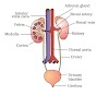
The nutrition in human beings takes place through Human Digestive System. It consists of alimentary canal and its associated glands. Various organs of Human digestive system in sequence are: Mouth, Oesophagus (or Food Pipe), Stomach, Small Intestine and Large
Intestine. The glands which are associated with this system are: Salivary glands, Liver and Pancreas. The human alimentary canal runs from mouth to anus and is about 9 metres long tube. The ducts of various glands open in to the alimentary canal and pour the secretions of the digestive juices in to the alimentary canal.
Now we will understand the various steps involved in the nutrition of human digestive system of human beings:
Now, the slightly digested food goes to stomach through food pipe i.e oesophagus. The walls of food pipe have muscles which can contract and expand alternately. When slightly digested food enters the food pipe, the walls started movements of contraction and expansion and this movement is known as peristaltic movement. And this peristaltic movement pushes food inside the stomach.
It consists of Mouth,Oesophagus and
Stomach,Small intenstine (Duodenum + Ileum) Large Intenstine (Colon)

It is 20 feet long.Villi provides more surface area (600 times) for better absorption.
At the place where small and large intenstine are joined, two organs are found.
1 .Caecum
2.Appendix
Food in the small intestine is called chyle.
The small intestine in human beings is the site of complete digestion of food like carbohydrates, proteins and fats.
1. The small intestine receives the secretions of two glands: Liver and pancreas. Liver secretes bile. Bile is a greenish yellow liquid made in the liver which is normally stored in the gall bladder. Bile is alkaline and contains salts which help to emulsify or break the fats or lipids present in the food. It makes the acidic food alkaline which comes from stomach so that pancreas can act on it and it also break the fats present in the food in to small globules making it easy for the enzymes to act and digest them. Pancreas is a large leaf like gland which lies parallel to and beneath the stomach. Pancreas secretes pancreatic juice which contains digestive enzymes like pancreatic amylase, trypsin and lipase. Amylase breaks down the starch, trypsin digests the proteins and lipase breaks down the emulsified fats.
2. The walls of small intestine contain glands which secrete intestinal juice. The intestinal juice contains a number of enzymes which complete the digestion of complex carbohydrates in to glucose, proteins in to amino acids and fats in to fatty acids and glycerol. Glucose, amino acids, fatty acids and glycerol are small, water soluble molecules. In this way, the process of digestion converts the large and small insoluble food molecules in to small, water soluble molecules. The chemical digestion of food is brought about by biological catalysts called enzymes.
3. After digestion the molecules of food becomes small and passes through small intestine and goes in to our blood. So, we can say that small intestine is the main region for the absorption of digested food. The inner surface of small intestine has millions of tiny, finger like projections called Villi which gives large surface area for absorption and the absorbed food goes in to our blood.
4. The blood carries digested and dissolved food to all the parts of the body where it becomes assimilated as part of the cell. This assimilated food is used by the body cells for obtaining energy as well as for growth and repair of the body. The undigested food stored in the liver in the form of carbohydrate called 'glycogen' and can be used by the body during requirement.
5. A part of the food which we eat cannot be digested by our body. This undigested food cannot be absorbed in the small intestine. So, undigested food passes from small intestine to large intestine. The walls of large intestine absorb most of the water from this food and become solid. Last part of the large intestine called 'rectum' stores this undigested food for some time and finally, egested from our body through anus as faeces or 'stool'. This process is known as egestion or defecation.

It is 5 feet long and 2-5 inches in diameter It has three parts :
Bacterial fermentation of indigestible materials.


Functions of Rennin or Chymosin or

Intestine. The glands which are associated with this system are: Salivary glands, Liver and Pancreas. The human alimentary canal runs from mouth to anus and is about 9 metres long tube. The ducts of various glands open in to the alimentary canal and pour the secretions of the digestive juices in to the alimentary canal.
Now we will understand the various steps involved in the nutrition of human digestive system of human beings:
Ingestion:
Food is ingested in human beings through the mouth and it is put in to the mouth with the help of hands.Digestion:
In the mouth itself the digestion of food begins in the mouth itself. Process of digestion is as follows: The mouth cavity or buccal cavity contains teeth, tongue and salivary glands. The teeth cut the food in to small pieces, chew and grind it. So, teeth help in the physical digestion. The salivary glands present in our mouth produce saliva and with the help of tongue saliva is mixed with food. As, we know that saliva is a watery liquid so it wets the food in our mouth and helps to swallow it easily. Many a times we have observed that when we see or eat a delicious food our mouth 'waters'. This is due to the saliva produce by the salivary glands. The salivary glands help in chemical digestion by secreting enzymes. Human saliva contains an enzyme known as salivary amylase which digests the starch present in food in to sugar. Thus, the digestion of starch or carbohydrate begins in the mouth itself. But food remains for very short time in mouth so, digestion of food remains incomplete in mouth.Now, the slightly digested food goes to stomach through food pipe i.e oesophagus. The walls of food pipe have muscles which can contract and expand alternately. When slightly digested food enters the food pipe, the walls started movements of contraction and expansion and this movement is known as peristaltic movement. And this peristaltic movement pushes food inside the stomach.
Parts of Human Digestive System
Small Intenstine
The small intestine is the largest part of the alimentary canal. It is known as small intestine because it is very narrow. The small intestine is arranged in the form of a coil in our belly.
1 .Caecum
2.Appendix
Food in the small intestine is called chyle.
The small intestine in human beings is the site of complete digestion of food like carbohydrates, proteins and fats.
Functions of Small Intestine
1. The small intestine receives the secretions of two glands: Liver and pancreas. Liver secretes bile. Bile is a greenish yellow liquid made in the liver which is normally stored in the gall bladder. Bile is alkaline and contains salts which help to emulsify or break the fats or lipids present in the food. It makes the acidic food alkaline which comes from stomach so that pancreas can act on it and it also break the fats present in the food in to small globules making it easy for the enzymes to act and digest them. Pancreas is a large leaf like gland which lies parallel to and beneath the stomach. Pancreas secretes pancreatic juice which contains digestive enzymes like pancreatic amylase, trypsin and lipase. Amylase breaks down the starch, trypsin digests the proteins and lipase breaks down the emulsified fats.
2. The walls of small intestine contain glands which secrete intestinal juice. The intestinal juice contains a number of enzymes which complete the digestion of complex carbohydrates in to glucose, proteins in to amino acids and fats in to fatty acids and glycerol. Glucose, amino acids, fatty acids and glycerol are small, water soluble molecules. In this way, the process of digestion converts the large and small insoluble food molecules in to small, water soluble molecules. The chemical digestion of food is brought about by biological catalysts called enzymes.
3. After digestion the molecules of food becomes small and passes through small intestine and goes in to our blood. So, we can say that small intestine is the main region for the absorption of digested food. The inner surface of small intestine has millions of tiny, finger like projections called Villi which gives large surface area for absorption and the absorbed food goes in to our blood.
4. The blood carries digested and dissolved food to all the parts of the body where it becomes assimilated as part of the cell. This assimilated food is used by the body cells for obtaining energy as well as for growth and repair of the body. The undigested food stored in the liver in the form of carbohydrate called 'glycogen' and can be used by the body during requirement.
5. A part of the food which we eat cannot be digested by our body. This undigested food cannot be absorbed in the small intestine. So, undigested food passes from small intestine to large intestine. The walls of large intestine absorb most of the water from this food and become solid. Last part of the large intestine called 'rectum' stores this undigested food for some time and finally, egested from our body through anus as faeces or 'stool'. This process is known as egestion or defecation.
Large Intenstine
1.Cecum:
Cecum or caecum, is a pouch or tubelike structure in the lower abdominal cavity that receives undigested food material from the small intestine and is considered to be the beginning of the large intestine.2.Colon:
It is thicker than small intestine and thinner than coecum.lt does not secret any enzymes. Digested residue is stored. It absorbs water from residue.3.Rectum:
It secrets no enzymes. It stores digestive residue and absorbs water from residue.lt is last part of the alimentary canal.Functions of the Large Intestine
The large intestine performs four major functions, included below:Absorption:
The absorption of water and mineral ions e.g., sodium and chloride. When the partially digested food reaches the large intestine, about 80 percent of the water from the food has been absorbed. The colon absorbs the rest of the water.Formation of faeces:
Formation and temporary storage of faeces. While the remnant food moves along the colon, it is mixed with mucus and bacteria, converted into faeces, and stored temporarily before being eliminated.Bacterial fermentation:
Bacterial fermentation of indigestible materials.
Maintaining the bacteria in the gut:
Maintaining a population of around 500 types of bacteria in the gut. The gut bacteria performs many useful functions other than fermenting undigested macronutrient material. The many functions of gut bacteria include interacting with the immune system, producing vitamins, stimulating the release of hormones involved in storage of fats as well as influencing our mood and feeling of well being.Digestion in Buccal Cavity
In buccal cavity, food gets mixed with saliva from salivary gland during chewing.
Composition of Saliva:-
99.4% water, 0.6% other compounds which includes inorganic (NaCl, KCI, Na2HP04, CaC03), mucous, salivary amylase or Ptyline, lysozyme. pH : 6.8
Total amount: 1-1.5 lit. With saliva, Pb, Hg and their iodides are excreted out. Secretion of saliva is under control of Medulla oblongata. Para sympathetic system increases secretion of saliva.Sympathetic system decreases secretion of saliva.
Functions of Saliva:-
i. To tie food particles.
ii. To make food moist and slimy.
iii. Lysozyme and Thiocyanate destroy bacteria.
iv. Ptyline converts starch into maltose, isomaltose and limit dextrine
v. Helpful in detecting taste.
vi. Helpful in water regulation.
Note: On feeling thirst, O.P. of body fluid is increased. Now food is like a ball called bolus which reaches stomach by peristalsis.
Digestion in Stomach
Spallanzani explained digestion in general and Beaumont explained it in man.As food comes in stomach, gastric juice is secreted by 350 lakh gastric glands. Gastric glands are stimulated by gastrin hormone secreted by G-cells of pyloric part.
Quantity of gastric juice becomes maximum in 1 to lh hours.
Composition of Gastric juice:-
99% water, 1% others which include inorganic salts, mucous, HCI (0.4%), pepsinogen, prorennin, gastric lipase, intrinsic factor.pH : 1 .0-1 .5. Total amount : 1-3 lit./24 hrs.
Functions of HCL:-
(i) To provide acidic medium.
(ii)To kill harmful bacteria.
(iii)To destroy living part of food.
(iv)To avoid rottening of food.
(v)To activate enzymes.
Pepsinogen ----------------> Pepsin
Prorennin -----------------> Rennin
Functions of Pepsin
pepsin
Protein -----------> Peptone + Proteoses
Functions of Rennin or Chymosin or
Curding enzyme
Renin
Caseinogen ---- --- Casein ------>
Paracasseinate
Functions of Gastric lipase
It is negligibly active.
Lipase
Fat > Fatty acids + Glycerol
Now food is called chyme, it comes in Duodenum. Here enterogastrone is secreted which stops digestion in stomach
Measures against Autodigestion of Stomach
There is a basic layer of mucous on mucosa.
Tight junction are present between cells.
Cells of mucosa are replaced at the interval of 2-3 days.
Note : A bacterium helicobactor pylori is responsible for peptic ulcer.
Digestion in Duodenum:-
It receives bile juice and pancreatic juice.
Hepatocrine stimulates synthesis of bile juice.
CCK stimulates contraction of gall bladder.
Secretin (discovered by Bayliss & Starling) and pancreozymine stimulate secretion of pancreatic juice.
First bile juice reacts on food by making the medium alkaline
Digestion in Small Intestine
Here food gets succus entericus stimulated by enterokrinine harmone.
Composition of succus entericus
98.5% water, 1 .5% others which includes mucous, inorganic salts, erapsin, maltase, isomaltase, limit Dextrinase, sucrase, lectase, lipase, polynucleotidase, phosphatase, nucleosidase
Erapsin
Tri/Di/Peptide ---------------
Aminopeptidase dipeptidase
Nucleotidase
Nucleotides ---------------> Pentose +
Nitrogenous base
Digestion of Cellulose:
ln man digestion of cellulose takes place in herbivorous. In rabbit it is not completed in one instance. Rabbit feeds on its evening faeces. It is coprophagy or pseudorumination. Such animals are coprophagous.
Food passes from alimentary canal two times, this phenomenon is dystrophy.
Absorption of Food- Absorption from Mouth
Here negligible tobacco, alcohol and some medicines as isoprenaline glyceryl tri-nitrate is absorbed.
Absorption from Stomach
Here glucose, water, alcohol and vitamin Here glucose, water, alcohol and vitamin B12 are absorbed.
Absorption from Intestine
Villi are helpful.
Vilicrinine stimulates it.
In Villi b.v. and lacteals present.
Carbohydrate and protein are absorbed by
b.v.
Lipid is absorbed by lacteals, colourless lymph becomes milky due to formation of chylomicrones. It reaches to heart first.
Carbohydrate and protein reach to liver.
Fructose is absorbed by facilitated Fructose is absorbed by facilitated diffusion.
Mannose and pentose by passive transport.
Glucose and galactose by active transport
1-120 by osmosis.
D.A.A. by passive transport.
L.A.A. by active transport.
Vitamins A and D by simple diffusion.
In columnar cells, acids phosphatase enzyme present, shows active process for absorption.
Egestion:
To give out undigested food from alimentary canal.
Faeces can remain in colon for 36 hrs then moving into rectum by gastro-colic reflex.
Faeces consists of 3/4 water and 1/4 solid.
In solid part, 3% bacteria, 30% roughage, 20% fat, 15% inorganic and 3% protein are present.
Dead mucosal cells, mucous, cholesteroal also present. NH3, CFI 4 are negligible.
Brown colour is due to stercobilin and stercobilinogen.
Foul smell is due to sketol, indol and treptophan.


No comments